
Clinical cases
Clinical cases: prostatic artery embolization
Three patient files treated for benign prostatic hyperplasia. The prostatic arteries were 3D reconstructed by our interventional radiologists, in order to concretely show what the preoperative scanner makes it possible to anticipate.
How to read these clinical cases
Before each prostatic embolization, a CT angiography maps the arterial network of the pelvis. Our interventional radiologists then produce a three-dimensional reconstruction from it, which makes visible what a simple cross-section does not show: the actual pathway of the prostatic arteries, their origin, and any potential communications with neighboring organs.
Why it is crucial
Prostatic arteries are small and their anatomy varies considerably from one man to another. Anticipating these variations before entering the operating room reduces procedure time, limits radiation exposure, and prevents non-target embolization.
The three cases below illustrate three distinct situations: standard anatomy, an arterial communication requiring protection, and a variant origin. In other words, three reasons never to approach an embolization without prior mapping.
What the CT angiogram concretely provides
The acquisition is performed after injection of contrast medium, in thin slices. The post-processing software then reconstructs the volume, which the radiologist rotates in space until the path of the prostatic arteries is isolated. This step takes a few minutes; it saves a lot of time in the operating room.
Three benefits derive directly from this. First, a reduction of procedural time the route is known, there is no need to look for it. Then, a reduction of radiation exposure and contrast volume, since exploratory acquisitions are becoming unnecessary. Finally, a prevention of non-target embolization, as communications with the bladder, rectum, or genital structures are identified prior to the injection.
The arterial anatomy of the prostate
The prostate is supplied primarily by branches originating from the inferior vesical artery. One usually distinguishes a central prostatic artery, which penetrates at the base of the gland near the bladder neck and descends along the prostatic urethra, and a peripheral prostatic artery, which runs along the lateral and posterior aspects before entering the gland.
To these two main trunks are added accessory contributions: the internal pudendal artery, the artery of the vas deferens, and the middle rectal artery. In the event of hypertrophy, this network develops considerably to supply blood to the'adenoma — which makes it all the more visible in imaging, but also all the more variable from one patient to another.
Why communications must be protected
The pelvic network is not partitioned. Anastomoses exist naturally between the prostatic arteries and neighboring territories: bladder, rectum, genital structures. As long as they remain patent, the microspheres injected can borrow them and embolize a non-targeted territory.
The principle behind the workaround is simple: block off communication before injecting coils, those small metal springs deployed inside the blood vessel. The area in question is thus put out of harm's way, and the embolization can proceed safely for it. This is precisely what the second case illustrates.
Clinical case 1
Standard anatomy
Male · 49 years old
Reason for consultation
- Frequent urination during the day
- Repeated night awakenings
- Weak urinary stream
Arterial mapping
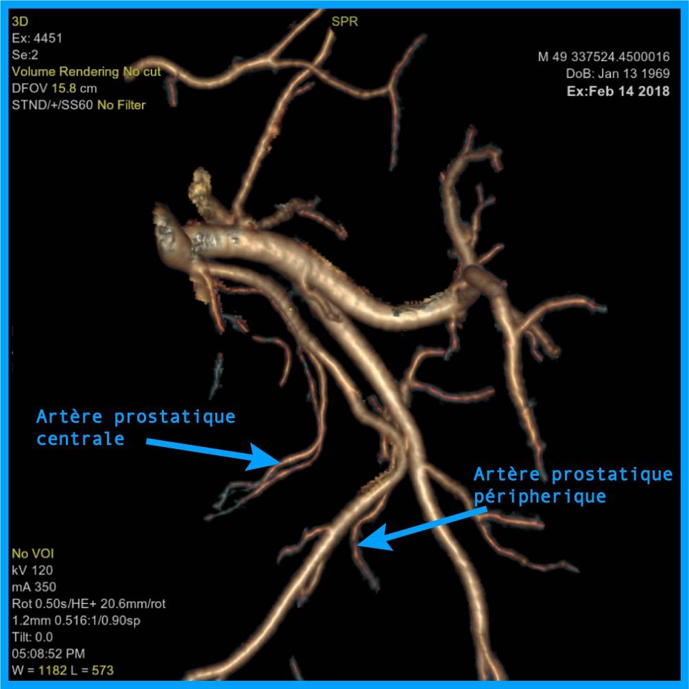
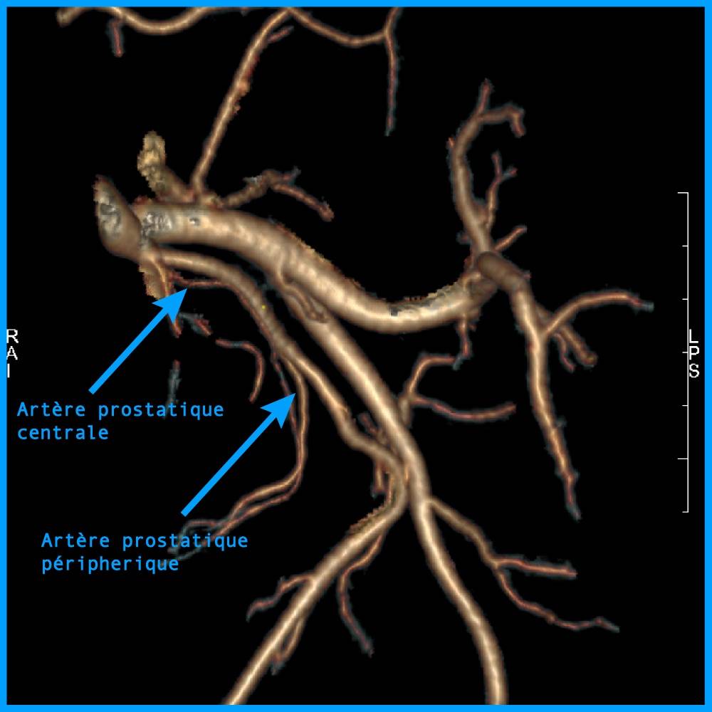
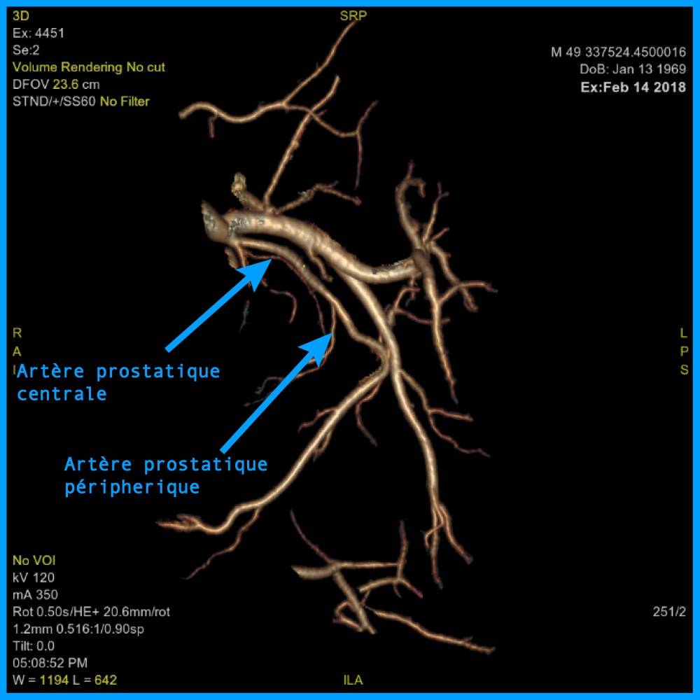
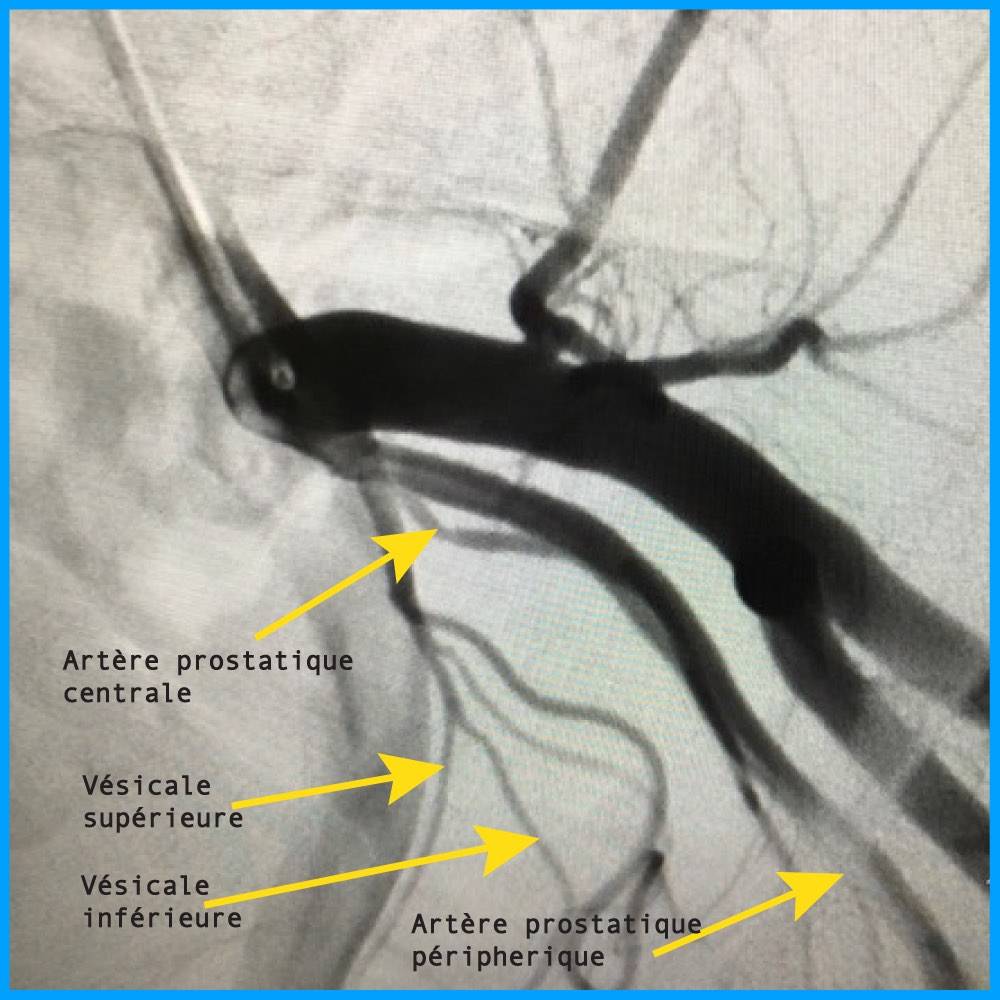
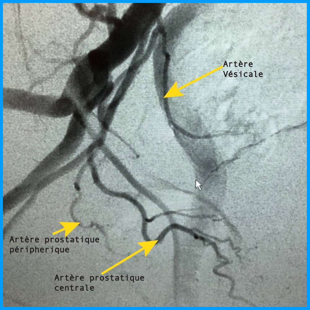
Prostatic arterial network of usual distribution, without notable communication with neighboring territories. 3D reconstruction allows the identification of the two prostatic arteries and planning of their catheterization.
The two trunks are clearly individualized: the central prostatic artery and the peripheral prostatic artery arise separately and follow their expected path.
Clinical case 2
Communication with the penile artery
Male · 65 years old
Reason for consultation
- Pollakiuria: very frequent urination
- Repeated night awakenings
- Weak urinary stream
Arterial mapping
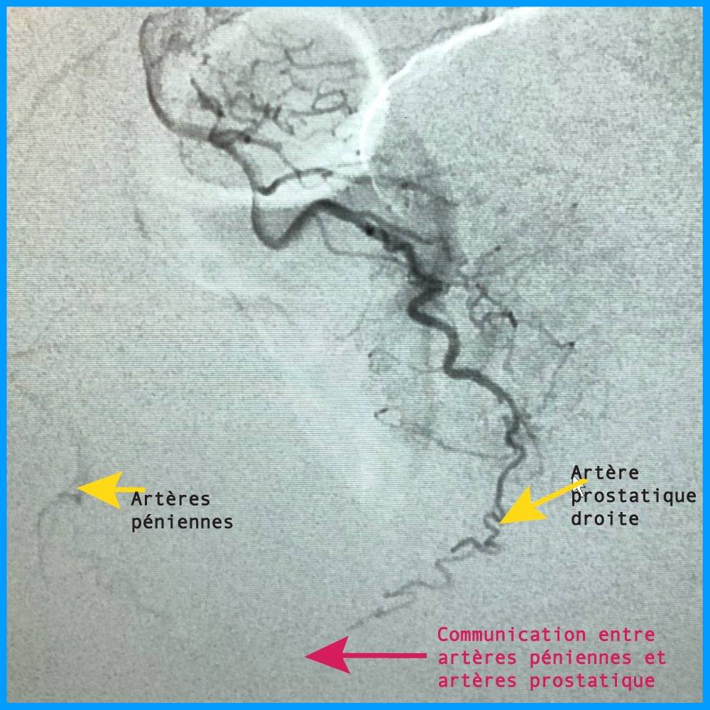
The CT scan highlights a communication between the prostatic artery and the penile artery. Without precautions, the injected microspheres could take this pathway.
Completed action — 2019 practice
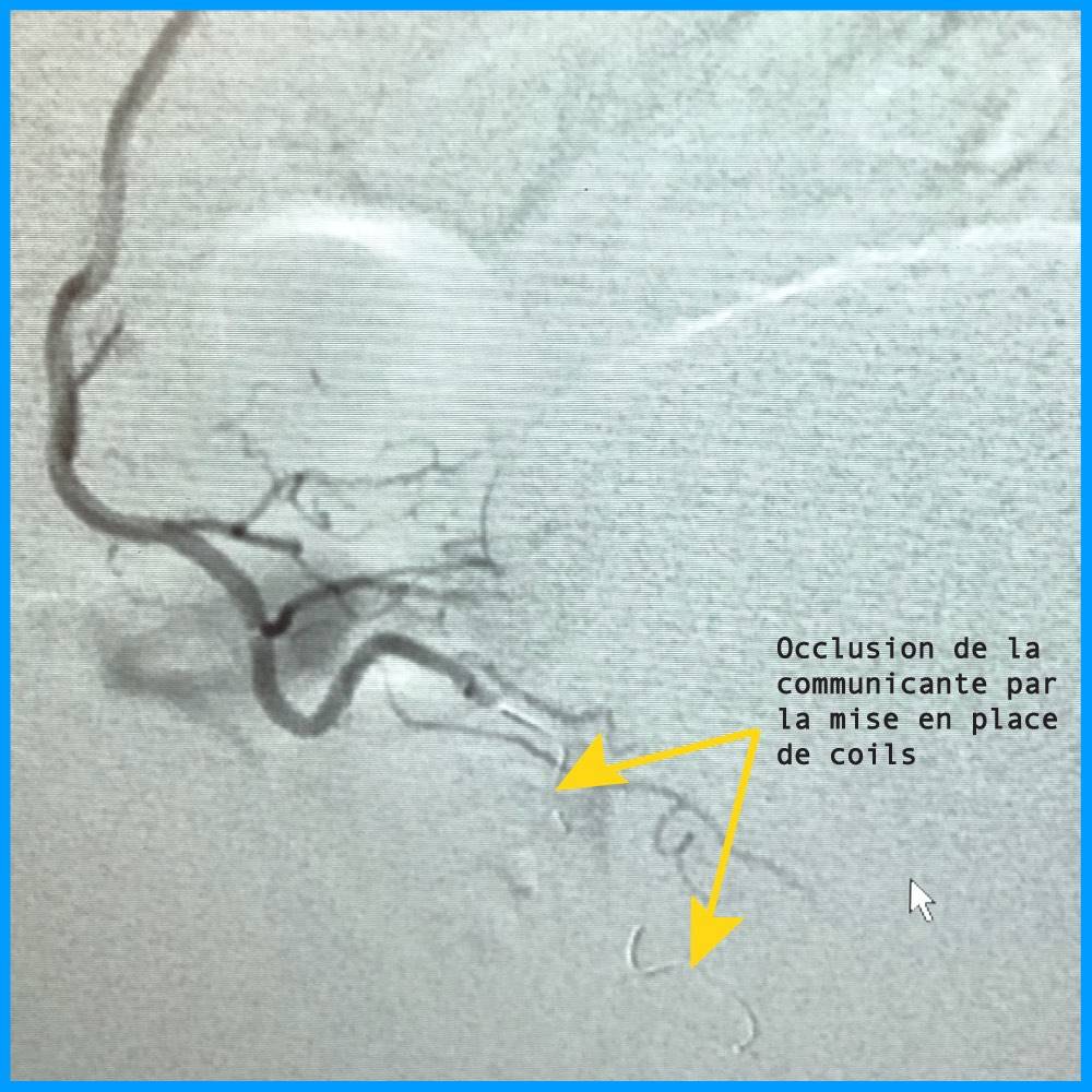
The communication was previously occluded by coils. Thus protected, the penile territory remains out of reach, and the microspheres are distributed exclusively in the prostate.
This anastomosis between the prostatic artery and the penile arteries is by no means exceptional: it is one of the anatomical variations regularly encountered. What matters is not its rarity, but the fact of identifying it before the injection rather than after. Unprotected, it would expose the patient to off-target embolization of the penile territory.
The coils are deployed in the communicating artery by the microcatheter, under radiological guidance. The control angiography then confirms two things: the communicating artery is occluded, and the prostatic arteries remain perfectly patent. Embolization only begins once this verification is done.
This case dates back to 2019 and documents a practice we no longer apply: today, we no longer use coils. The evolution of our technique allows us to manage these communications differently. The case is still presented here because it perfectly illustrates the challenge: identifying an anastomosis before injecting.

Clinical case 3
Anatomical variant of origin
Male · 59 years old
Reason for consultation
- Frequent urination
- Weak urinary stream
- Pain
Arterial mapping
Original variant: the prostatic artery arises from the gluteal artery and not from its usual origin. A configuration that, if not identified beforehand, considerably lengthens the search in the operating room.
Importance of mapping
Identified before the procedure, this variant was addressed directly. This is precisely the type of situation where the preoperative scan makes the difference between a smooth procedure and a catheterization failure.
In the usual arrangement, the prostatic artery arises from the inferior vesical artery, which itself branches from the anterior trunk of the internal iliac artery. Here, the central prostatic artery originates from the gluteal artery, that is to say from a posterior trunk. An operator unaware of this will look for its origin where it is not found.
The consequences of such blind research are concrete: prolonged procedure time, additional radiation, a larger volume of contrast agent, and sometimes outright failure of the catheterization. This is one of the reasons why patients are referred to us after an unsuccessful first attempt elsewhere.


What these cases demonstrate
These three cases, chosen from thousands of proceedings, have one thing in common: in each of them, the technical decision was made prior to the intervention, based on the imaging.
- Arterial anatomy is never standard — variable origin, communications with the bladder, rectum, or genital structures, very fine calibers
- An identified communication is a manageable communication — prior coil embolization protects adjacent territories
- An anticipated variant saves time — therefore reduces radiation and the amount of contrast agent
This also explains why the operator's experience carries so much weight in this technique. Recognizing a variant, deciding to protect a territory, achieving distal and complete embolization: these skills are acquired through volume.
Three technical questions that these cases raise
What is called an off-target embolization? This is the migration of microspheres into an area other than the prostate—bladder, rectum, genital structures—via an unidentified arterial communication. It may result in transient local symptoms. Its prevention relies entirely on prior imaging and monitoring of distribution during injection.
What are coils used for, and why don't we use them anymore? These are fine metal coils deployed in a vessel to occlude it in a targeted manner. They do not treat the adenoma: they protect a neighboring territory by closing off the pathway that the microspheres could take. We no longer use coils in our center. The evolution of our technique now allows us to do without it. The images of the second case, taken in 2019, therefore document a practice that we have since evolved.
Why a three-dimensional reconstruction? Because a prostatic artery measures less than a millimeter and follows a tortuous path in all three planes of space. On an isolated slice, it appears for only a few millimeters. Reconstructed in volume and manipulable, it becomes legible along its entire path, from its origin to its penetration into the gland.
Is your anatomy compatible?
Send your file and test results. We will tell you what arterial mapping can make possible in your case.
Frequently asked questions
Are these cases representative of all patients?
No, and that is precisely why they are presented. Each illustrates a different anatomical configuration. They show the variability of the prostatic arterial network, not a standard result. Your own anatomy will be evaluated by a angioscanner dedicated.
Is the preoperative CT scan routine?
It is performed in the vast majority of cases prior to prostatic embolization. It is particularly recommended in patients with marked atherosclerosis or tortuous vessels, in whom catheterization may prove difficult.
Does an anatomical variant prevent embolization?
Rarely. An identified variant is an affordable variant: it modifies the catheterization strategy, not the feasibility of the procedure. Truly blocking situations are more related to the general condition of the arteries than to their layout.
What happens if a communication is discovered during the intervention?
It is occluded before proceeding, as in the second case presented. The advantage of prior mapping is precisely to anticipate it: discovered during the procedure, it lengthens the procedure and requires real-time adaptation.
Can I send my own scan for review?
Yes. If you already have a CT angiography or pelvic imaging, please send it: it will be reviewed and will allow an initial assessment of your arterial network even before the consultation.
Get your application evaluated
Send your urinalysis, your ultrasound, your latest PSA or your scanner if you have one. We will tell you what is missing and whether embolization is an option in your situation.
Prostate Center — Blomet Clinic
136 bis rue Blomet, 75015 Paris · Monday to Saturday, 9 AM – 6 PM · +33 6 32 84 14 31
Related pages
Editorial responsibility. Medical content written and validated under the responsibility of Dr. Antoine Hakimé, interventional radiologist. Last medical review: September 2026.
Privacy. The presented cases are anonymized: no data allowing the identification of a patient is published. The images are published with the consent of the patients concerned.
Information. These cases illustrate situations encountered in our practice. They in no way prejudge the individual outcome: each anatomy and each indication requires a specific evaluation.